These pages describe our research project on Fast Field-Cycling Magnetic Resonance Imaging.
This research is being carried out in the School of Medicine, Medical Sciences and Nutrition at the University of Aberdeen , in the North-East of Scotland.
We are associated with the Aberdeen Biomedical Imaging Centre .
- Overview
-
Aberdeen was lead partner in the EU-funded Horizon-2020 research project "IDentIFY" (Improving Diagnosis by Fast Field-Cycling MRI) , which ran for four years from January 2016.
Our FFC-MRI project involves making a step change in magnetic resonance imaging (MRI) technology, by breaking one of its fundamental "laws" - that the applied magnetic field must be held constant during image acquisition. By deliberately switching the magnetic field during the collection of MR images, we are able to gain access to radically new types of endogenous contrast.
The project is generating the enabling technology to make FFC-MRI an invaluable tool for basic biomedical research, through to clinical research and diagnosis. We are also heavily involved with projects to investigate biological and medical applications of FFC-MRI.
The concept of FFC-MRI is explained in this animation:
In this video, Prof. David Lurie and team discuss their work:
- About
-
Rationale Behind Field-Cycling
Much of the "contrast" in MR images arises from differences in the T1 relaxation time between normal and diseased tissue. For example, tumours normally exhibit significantly longer T1 values than their surrounding normal tissue, making them stand out in MR images.
Studies over the years on small tissue samples have shown that the way in which a tissue's T1 varies with magnetic field could also be a potential indicator of disease. However, that information is completely invisible to conventional MRI scanners, because each scanner can only operate at its own magnetic field (e.g. 1.5 T or 3.0 T).
The aim of our project on "Fast Field-Cycling MRI" (FFC-MRI) is to build and use new, very special types of MRI scanner in which the magnetic field can be switched (or "cycled") during the scan, so that the pattern of how T1 varies with magnetic field can be measured. This new type of MRI should be even more sensitive to changes in tissues brought about by disease, potentially allowing earlier and more reliable diagnosis of some medical conditions.
We have built a range of FFC-MRI scanners which have allowed us to perform relaxometry studies and field-cycled MRI measurements of large samples, as well as human volunteers and patients. Our equipment can be seen on the Facilities panel below.
Fast Field-Cycling NMR Relaxometry
Fast Field-Cycling Nuclear Magnetic Resonance (NMR) has been used for decades in order to measure the variation of T1 with magnetic field strength of small samples. A field-cycling pulse sequence comprises three distinct periods, called "polarisation", "evolution" and "detection", each applied at a different magnetic field strength. The magnetic field during the evolution period can be changed, but the field applied during the detection period is always the same, so that the NMR signal is always at the same frequency.
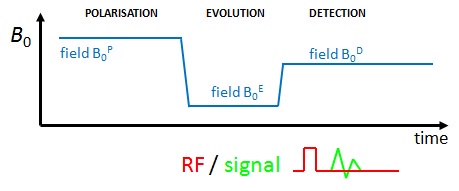
Field-cycling pulse sequence, showing the three periods. Relaxation takes place during the Evolution period, while signal detection occurs during the Detection period. A field-cycling relaxometry experiment involves repeating a T1-measurement pulse sequence - e.g. Inversion Recovery - each repetition using the same Detection field but a different Evolution field strength. The result of the experiment is a set of NMR signals obtained after the spins have "evolved" at a range of field strengths, and these can be analysed to produce a graph of T1 versus field strength (the T1 dispersion curve). The diagram below shows an example of a field-cycling relaxometry pulse sequence, which has been adapted for MRI by the addition of magnetic field gradients during the detection period.
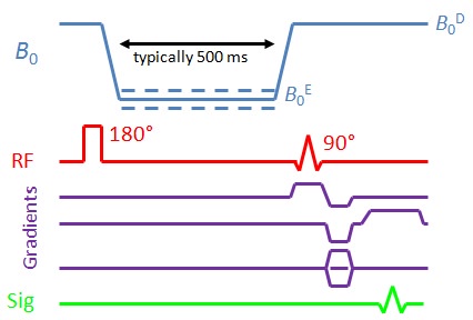
Field-Cycling Inversion-Recovery pulse sequence for relaxometric imaging measurements Quadrupole Dips
Whereas one might expect to see a gradual, smooth increase in T1 with field strength, with biological samples this increase is often superimposed by three pronounced "dips" where the T1 value drops by 10 to 15% over a small range of field strengths. These are known as "quadrupole dips" and are found at well-defined field strengths (16 mT, 49 mT and 65 mT) where the Nitrogen-14 nuclear quadrupole resonance (NQR) and the proton NMR frequencies are equal. T1 is reduced at these field strengths because the nuclear spins of hydrogen atoms on the "backbone" of protein molecules are strongly coupled to the nuclear spins of adjacent nitrogen atoms, and this enhances the efficiency of proton NMR relaxation at the NQR frequencies.

T1 dispersion curve measured from a volunteer's forearm. Quadrupole dips centred on 49 mT and 65 mT can clearly be seen. In fact, the "depth" of the quadrupole dips provides information about the concentration of protein in the sample under study, and we are investigating the possibility of protein imaging by "quadrupole-dip" MRI. By implementing a field-cycling inversion-recovery imaging pulse sequence, it is possible to collect images "on" and "off" a quadrupole dip. In areas of the body with large protein concentrations these will exhibit markedly different signal intensity. This is demonstrated in the images shown below of a volunteer's thighs, obtained with the evolution field set on- and off- the quadrupole dip at 65 mT. Muscle regions appear significantly darker (i.e. lower NMR signal) in the 75 mT image compared to the 65 mT image, due to the differences in T1 of the tissue at the two field strengths.
FC-IR images of a volunteer's thighs, obtained at evolution fields of 65 mT 
FC-IR images of a volunteer's thighs, obtained at evolution fields of 75 mT - Facilities
-
We have developed novel equipment to carry out our research on fast field-cycling MRI (FFC-MRI). Many of these MR systems are unique in the world, and offer unprecedented facilities for research, and for their use in applications. The techniques themselves are described elsewhere on this site.
In our first-generation whole-body field-cycling MRI system (shown below), the detection magnetic field was provided by a permanent magnet which generated a vertical field of 59 mT. Just inside the bore of the permanent magnet was a resistive magnet, made from copper conductor in a saddle configuration. When electric current was sent through the resistive magnet windings they generated a field of up to 59 mT, in opposition to the permanent magnet's field. In this way, switching on and off the current switched the total field in the centre of the magnet down and up in value, and the precise field value could be selected by choosing the correct current to send through the resistive magnet coils.
The typical switching time between magnetic field levels was 20-30 ms, significantly shorter than the T1 relaxation times found in most biological samples or in the human body.

The original 59 mT whole-body FFC-MRI system (with its copper RF screening removed)More recently, we have built a whole-body FFC-MRI scanner based on a single-magnet design. The use of a single resistive magnet makes the pulse sequence more flexible, as the detection field can be altered as well as the polarisation and evolution magnetic fields. However, this flexibility comes at the expense of potential instability of the magnetic field during the detection period. Therefore, particular attention must be paid to the design and implementation of the control circuitry and the magnet power supplies.

0.2 T whole-body magnet for FFC-MRI 
0.2 T FFC-MRI system, including 3-axis square Helmoltz coils for environmental field correction 
Service end of 0.2 T FFC-MRI magnet, showing water and electrical connections 
Power supply cabinets (3 x 650 A) for 0.2 T FFC-MRI scanner (International Electric Co., Finland) In addition to the home-built scanners described above, we have a commercial bench-top field-cycling relaxometer (Stelar SMARtracer), capable of making T1 dispersion measurements from 10 kHz to 10 MHz proton Larmor frequency on small (~1 ml) samples.

Our lab's benchtop field-cycling relaxometer - Funding
-
Funding for the FFC-MRI project has come from a number of sources, including the following:
- 2004-06 Leverhulme Trust grant (PI D.J. Lurie), "Field-Cycled Magnetic Resonance Imaging", £82k.
- 2007-11 RCUK/EPSRC Basic Technology grant (PI D.J. Lurie), "Fast Field-Cycling Magnetic Resonance Imaging ", £2.42m.
- 2011-13 Arthritis Research UK grant (PI G.P. Ashcroft), "Assessment of Fast Field-Cycling MRI for the Imaging of Articular Cartilage and Osteoarthritis", £191k.
- 2012-13 EPSRC Follow-on Fund grant (PI D.J. Lurie), "Field-Cycling Add-on for Clinical MRI Scanners ", £200k.
- 2013-16 EPSRC grant (PI D.J. Lurie), "Zero-Field MRI to Enhance Diagnosis of Neurodegeneration ", £979k.
- 2016-19 EU Horizon 2020 grant (PI D.J. Lurie), "Improving Diagnosis by Fast Field-Cycling MRI ("IDentIFY") ", €6.6m. Project was led by Aberdeen and included 9 partners in 6 countries. The IDentIFY project webpage is here .
- 2019-20 EU H-2020 ATTRACT 3rd-party grant (PI S. Geninatti-Crich, Turin University), "Monitoring Tissue Implants by Field-Cycling MRI of Quadrupolar-Peak Contrast Agents (QP-MRI)", €100k.
- 2020-23 British Heart Foundation (PI D. Dawson), "The Next Leap in Cardiac MRI: Cycling the Field ", £277k.
- 2020-22 Chief Scientist Office (Scottish Government) (PI M.-J. MacLeod), "Proving the Utility of Fast Field Cycling MRI in stroke and small vessel disease (PUFFINS) ", £299k.
- 2021 Sir Jules Thorn Charitable Trust (PI D.J. Lurie), Medically-related donation : "Power supply unit for new FFC-MRI scanner", £270k.
Our current and recent funders include:

British Heart Foundation 
Chief Scientist Office

EPSRC 
Arthritis Research UK

European Commision

The SIr Jules Thorn Our industrial collaborators include:

Stelar 
IECO - People
-
Core Research Group
-
Ms Amnah Alamri
Research Postgraduate Student

Dr Lionel Broche
FFC team leader
The Hall Family Lectureship in Medical Physics
Dr Gareth Davies
Research Fellow
Professor David Lurie
Emeritus Professor
Dr Vasiliki Mallikourti
Research Fellow

Dr James Ross
Lecturer

Mr Robert Stormont
Research Postgraduate Student (part-time)
Local Research Collaborators
- Mr Pragnesh Bhatt
Honorary Clinical Senior Lecturer
- Dr Tanja Gagliardi
Honorary Senior Clinical Lecturer
- Dr German Guzman Gutierrez
Honorary Associate
- Dr Mary Joan MacLeod
Senior Clinical Lecturer
- Dr Arnab Rana
Honorary Senior Clinical Lecturer
- Dr Leslie Samuel
Honorary Clinical Senior Lecturer

Mr George Ashcroft
Senior Clinical Lecturer

Professor Dana Dawson
Chair in Cardiovascular Medicine

Mr Yazan Masannat
Consultant Oncoplastic Breast Surgeon
and Honorary Clinical Senior Lecturer
Dr Nicola Mutch
Reader in Applied Medicine
Dr Justin Rochford
Reader
Dr Justin Rochford
Reader
Former PhD Students on FFC-related projects
- Dr Marcello Alecci
- Dr Chang-Hoon Choi
- Dr Dara Ó hÓgáin
- Dr Ian Nicholson
- Dr. Nicholas Payne
- Dr Kerrin Pine
- Dr Chittakorn Polyon
- Dr Patana Puwanich
- Dr Fraser Robb
- Dr James Ross
- Dr David Yeung
- Dr Wiwat Youngdee
- Dr Vasileios Zampetoulas
- Dr. Nicholas Payne
-
- Public Engagement and Publicity
-
Here you can find information about recent activities aimed at informing the public about our research. You can also find details of the public engagement activities that took place during our EU Horizon-2020 project "IDentIFY"
General Engagements
British Science Week
Cafe Scientifique Talk
- Publicity
-
TV and Radio
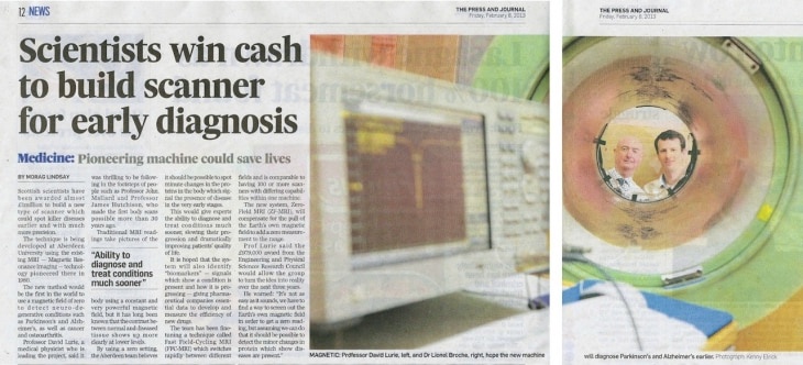
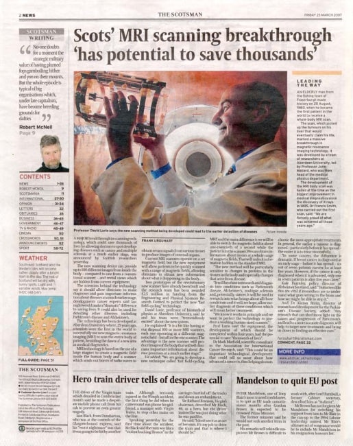
News Articles
Online articles
Radiology "Trade" Articles
General-Interest Articles
Scientific Publications


Biomedical Physics Building University of Aberdeen Foresterhill Aberdeen AB25 2ZD United Kingdom