The FPR2 Ligand W-peptide: a new therapeutic approach to treat Atherosclerosis
For my Hotstart internship, I worked under Dr Dawn Thompson's supervision for nine weeks. The lab-based project investigated the formyl peptide receptor 2 (FPR2), a G-protein coupled receptor which promotes tissue healing.
Ligands such as SAA, annexin-1, resolvin D1 and lipoxin A4 bind to formyl peptide receptors and activate downstream signalling that can either contribute to inflammation or resolution of inflammation. These work as a positive loop in order to maintain homeostasis. Failure of this process can induce diseases such as atherosclerosis and obstructive pulmonary disease.
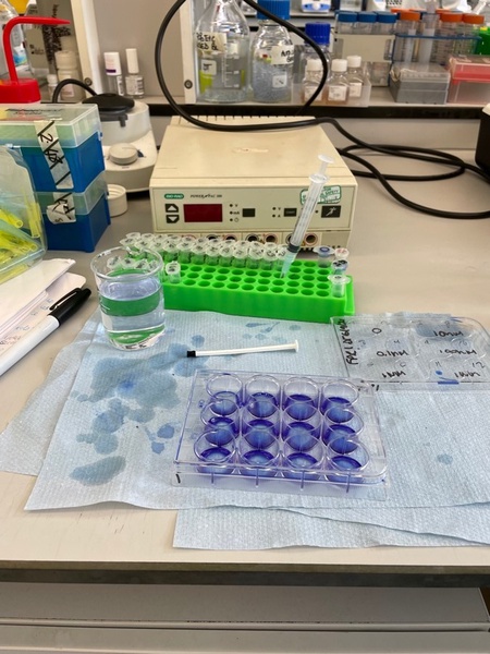
I had the opportunity to learn different techniques such as Western blotting, cell culture and sample preparation. Two different experiments were conducted with HEK293 cells expressing either FRP2 or FPR1 and mice liver tissue.
A cell stimulation experiment using W-peptide (a synthetic agonist) was conducted on HEK293 cells. Different concentrations of the drug were added to the cells and gels were produced to analyse the relative activation of the receptor with increasing drug concentration. Lastly, after knowing the exact concentration needed to activate the receptor, we conducted a time-response test.
Experiments showed that W-peptide is an agonist for both FPR1 and FPR2, although FPR1 relative activation is milder comparted to FPR2 relative activation. FPR2 reached its maximum activation at 100nM, therefore, the time-response experiment was carried out using this drug concentration. Results from the latter showed that FPR2 reaches its maximum activation at 10 minutes.
The second type of experiment was done with liver tissue samples from female ApoE-/- mice, a model of atherosclerosis, fed a high fat/high cholesterol diet (to accelerate plaque formation) injected with either saline or W-peptide. Western blots were incubated with pERK, Pp38 or BIP primary antibodies and reprobed with either Tubulin or GAPDH in order to confirm the protein loading in each well of the gel.
During the first experiments, we observed two bands when reprobing BIP membranes using mouse GAPDH, this suggested that mouse GAPDH could possibly be binding to IgG in the liver samples. Therefore, we ran an experiment where we ran two gels and probed transferred membranes with either anti-rabbit or anti-mouse total ERK followed by secondary HRP-conjugated antibody or two that were probed only with rabbit or mouse secondary antibody. The results showed that anti-mouse HRP detects IgG in mice liver samples, therefore, not a good option for these experiments. Hence, GAPDH antibody raised in rabbit was used instead.
This internship was a really good learning experience that allowed me to learn useful techniques which will be essential for my honours project and further studies.


