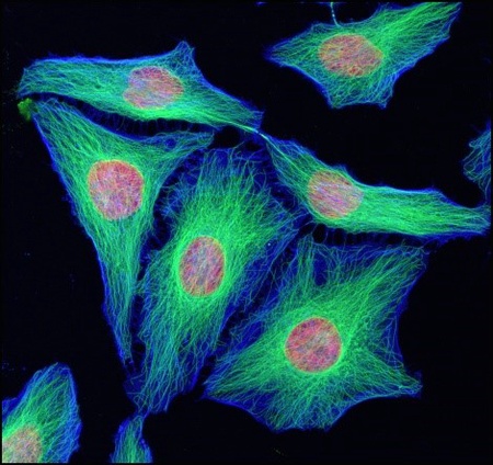
Microscopy plays an important part in everyday research. The University’s Microscopy and Histology Facility provides researchers on both campuses with access to advanced technologies.
Originally established in 2004 as two separate facilities (Histology & Electron Microscopy and Imaging), the two were merged in 2010 to form the service that exists today. Located within the Institute of Medical Sciences, we welcome users from all disciplines - whether you are a member of staff, a student, or a visiting scientist from another institution, we’re here to help whatever your level of expertise.
Our skilled staff are on hand to help you get the most from a wide range of specialist microscopy services and resources, including sample preparation and sectioning for light and electron microscopy, slide scanning, live cell imaging, fluorescent microscopy, and Micro CT.
We have a wide range of modern equipment including 18 different types of microscope. Our most recent acquisition is a Zeiss LSM880 Airyscan confocal microscope funded by the Wellcome Trust. We look at a diverse range of samples including bone, tissue, cells, bacteria, nanoparticles, and even insects. Bread, seaweed and sea water plankton are just some of the more unusual samples to pass through the lab. Throughout the year we run the Microscopy course for new users. This takes place over 2-3 days and includes a series of talks and demonstrations covering all aspects of microscopy and histology. We have also run a specialised course in Immunofluorescence.
We are actively involved in University outreach activities – such as Techfest, Mayfest, Doors Open day – and with work experience placements for secondary school pupils. We are also involved in the Scottish Microscopy Society. Established in 1968, the group aims to bring microscopists from all disciplines together to share ideas, methods, and new technologies. We were recently invited by St Andrews Photographic Society to give a talk on how ‘Photography Under The Microscope’ has changed over the years. On the back of this, a talk to Dundee Photographic Society is planned for next year. You can view some of the Facility’s award winning images on the Wellcome Collection and Science Photo Library websites.
In collaboration with the University’s Toolkit team, we have produced a series of short introductory videos on Light Microscopy, Fluorescent Microscopy and Electron Microscopy, and also a short instructional video on how to set up a microscope for Brightfield imaging.
We welcome new users – whether to help you get started with a new study, or to help you sort out an unexpected result, we are always up for a challenge!
Contact us at microscopy@abdn.ac.uk to arrange a visit/appointment.
Kevin Mackenzie, Microscopy Facility Manager
Facebook
@AberdeenMicro
Instagram
@aberdeen_microscopy
Twitter
@Abdn_Micro


