Women from Aberdeen and Aberdeenshire are being sought to help in the testing and calibration of a new type of MRI scanner, which may provide doctors with more reliable information about breast disease.
Researchers need to understand how healthy breast tissue looks to the new scanner, helping them to correctly identify indications of disease in future scans of patients.
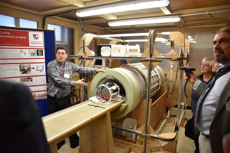
The new machine, called “Fast Field-Cycling MRI” or FFC-MRI, uses a weak, switchable magnetic field along with pulses of radiowaves to generate data about subtle changes in the body’s tissues.
It will be especially useful to see how the appearance of breast tissues changes during the menstrual cycle, using the FFC-MRI scanner.
By studying healthy participants, the team will develop a very clear data profile for normal breast tissue. This will make it easy in the future to observe the earliest indications of disease.
The team hope that this will lead to earlier diagnosis of breast cancer, potentially allowing clinical intervention to begin sooner.
This new technology offers a very real chance to improve the outcomes of future breast cancer patients. But the team do require a number of willing volunteers to donate their time at this important stage.
Professor Lurie said: “We’d love to welcome volunteers to our centre. Participants will play a vital role in progressing this new technology, by helping us understand more precisely the markers of healthy breast tissue.
“Volunteers will be able to get a first look at some exciting new technology that won’t be commercially available for years yet, and they will help Aberdeen lead on this important work, which we hope will save the lives of women. The process is entirely safe and should take no more than an hour of each volunteer’s time.”
The new scanner has been developed in part through a 4-year, EU-funded project called “IDentIFY”, standing for “Improving Diagnosis by Fast Field-Cycling MRI”. Led by the University of Aberdeen, the research involves partners from 9 sites across Europe, funded by a grant of €6.6M from the EU’s Horizon-2020 programme.
Aberdeen has always led the world in Magnetic Resonance Imaging (MRI). The first-ever full-body MRI machine was developed in Aberdeen during the 1970s. The first patient was a man from Fraserburgh, who was successfully scanned in 1980. That first scanner, the “Mark-I” is now part of an exhibition open to the public at the Suttie Arts Space in Foresterhill. Since that breakthrough 39 years ago, MRI machines have spread across the world, becoming a vital diagnostic tool in every major hospital.
Now Aberdeen is once again at the global vanguard in the development of a new type of scanner. Under Professor David Lurie, a team at the University of Aberdeen has led a European-wide research project aimed at improving the FFC-MRI scanners and finding out how they can benefit doctors and patients. The machine works at low magnetic fields to reveal new information about the body’s tissues that is invisible to current medical scanning equipment.
The team are currently looking for healthy female volunteers to undergo three separate breast scans throughout their menstrual cycle. Volunteers must be aged 16 and over and have a regular and stable menstrual cycle. Volunteers must be premenopausal and must not take the contraceptive pill or any other form of hormone therapy. Potential participants are encouraged to contact Ms. Stacey Dawson, the project’s Clinical Studies Coordinator, by emailing stacey.dawson@abdn.ac.uk.


