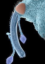
An image of a bat’s tongue may not sound like the most likely art exhibit. The same could be said about pictures of mildew, nerve cells and the head of the fearsome Scottish biting midge.
But add some colour and these images - taken with microscopes - become really rather striking.
All these and more, from a world not normally seen by the naked eye, go on show to the public tomorrow (Friday, January 25) at the Art Gallery within Aberdeen Royal Infirmary.
Micro art is an exhibition of microscope images – or micrographs - captured and manipulated by the University of Aberdeen's Kevin Mackenzie.
The images have been taken using various microscopes, and then cleverly infused with colour.
It's unlikely that visitors to the exhibition will have seen skin cells, an aphid, a blood clot, and rust spores – also among the 30 images on show – depicted in this way.

"There seems to be a natural fascination for all things microscopic. I'm hoping visitors will enjoy seeing the world from a different perspective – one not normally seen without the aid of a microscope."
The free exhibition, organised by the Grampian Hospitals Art Trust in association with the University, runs from January 25 to March 7.
Visit http://www.abdn.ac.uk/emunit/microart/gallery/index.html


