A centuries old theory that has long been fiercely contested has now been backed-up by UK scientists.
The theory first mooted in the late 1800s, suggested that paired fins evolved from gills, but has since been widely discounted. Now researchers have found evidence to support the original hypothesis.
It was previously believed that comparative anatomist Carl Gegenbaur’s theory that paired fins evolved from gill arches couldn’t be possible as these structures develop from two distinctly different sets of cells in the embryo. Fins come from a population of cells called mesoderm while the gill arches develop from a population called neural crest.
However, new research published today in eLife led by Dr Victoria Sleight, now at the University of Aberdeen, alongside Dr Andrew Gillis from the University of Cambridge supports the hypothesis from the 1800s.
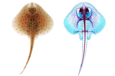
By studying embryos of the skate, a cartilaginous fish closely related to sharks, Dr Sleight and Dr Gillis found that gill arches develop from both neural crest and mesodermal cells – what is known as dual origin.
Dr Sleight commented: “Although the distinct embryonic origin of the gill arches and paired fin skeleton is widely assumed, it has never been formally tested in fishes. Understanding the embryonic origin of gill arches and paired fins sheds light on their possible evolutionary relationship.”
“We labelled neural crest and mesoderm cells of skate embryos by microinjecting them with fluorescent dyes, and we followed these cells over several weeks of development to discover the skeletal element to which they contributed. Fishes possess a series of gills that sit behind the jaw, and we found that the skeleton of the jaw and first gill were formed entirely from neural crest cells. However, surprisingly, the skeleton of the rest of the gills in the series were formed from a mixture of neural crest and mesoderm cells, and the fins were formed entirely from mesoderm. This discovery reveals a previously unrecognised embryonic continuity between gill and fin skeletons, and explains the anatomical similarities that led Gegenbaur to propose an evolutionary relationship between these structures nearly 150 years ago.”
Dr Gillis added: “I think this work, and the classical studies that inspired it, illustrate different ways of thinking about evolutionary change. From an anatomical perspective, we can look at two structures that loosely resemble one another, like the skeletons of gills and fins, and speculate that one might have undergone an evolutionary transformation into the other. But by studying embryonic development, we can gain insight into why and how these two structures resemble one another.
“It has long been thought that similarities between the gill and fin skeletons of fishes are merely coincidental. Our finding that these structures develop from a common pool of cells suggests otherwise – that gills and fins share a much deeper and more substantial evolutionary relationship that is reflected in their embryonic development.”
The team conducted much of their research at the Marine Biological Laboratory in Woods Hole Massachusetts, over several years.
Dr Sleight and Dr Gillis’ work was supported by a research grant from the Leverhulme Trust, and by a Royal Society University Research Fellowship, a Junior Research Fellowship from Wolfson College and a Whitman Early Career Fellowship from the Marine Biology Laboratory.


