Parastomal Hernias
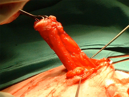
Operative photograph of local stomal hernia repair. The stoma is being raised from surrounding abdominal wall.
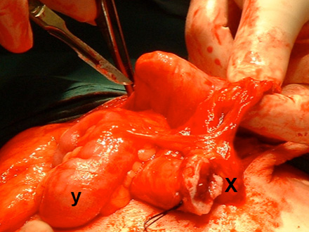
Operative photograph of the same patient. The surgeon's finger is in the hernial sac. X marks the stomal orifice and y marks the small intestine, which was in the sac.
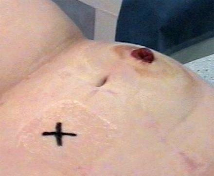
Operative photograph showing stoma site being marked prior to resiting.
A hernia is defined as a protrusion of a viscus or part of a viscus through a defect, which is congenital or acquired.
Therefore a parastomal hernia is produced by the heniation of small intestine and occasionally large intestine through the defect in the abdominal wall through which the stoma passes.
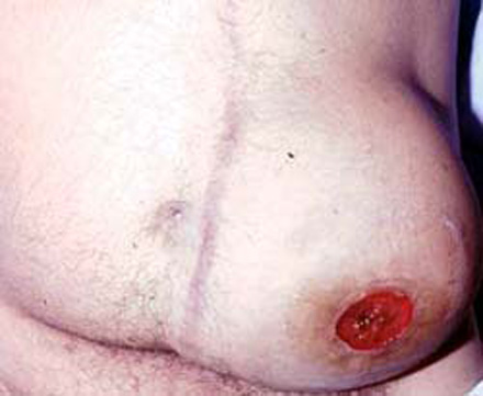
Large parastomal (colostomy) hernia. Note the large bulge which makes it difficult to fit the bag. This will lead to leakage of contents and excoriation of the surrounding skin.
N.B. Parastomal hernias are very common and the incidence increases with time. Surgeons have tried many techniques to prevent this complication with mixed results. Most surgeons recommend that a colostomy or ileostomy is brought through the rectus abdominus and not along the lateral edge of the rectus sheath.
Problematic parastomal hernia
The main complications of a parastomal hernia are;
- Inability of bag to remain adherent
- Leakage and excoriation
- Intestinal obstruction
When the hernia produces significant problems, then surgery is required. This may involve:
- Local repair
- Mixed results (high recurrence rates)
- Technically challenging
- No laparotomy needed
- Resiting of stoma
- Moved to opposite side
- Involves laparotomy
- Complex surgery
