Basic Interpretation
There are 3 main parts to the ECG waveform.
- P-wave
- QRS complex
- T-wave
When all of these occur in the correct order with the correct time interval in between them, this is called sinus rhythm. All the examples we are looking at on this page are using the readings from limb lead II.
Atrial Depolarisation
- The SA node discharges an action potential
- This flows through special fibres and causes the atria to depolarise/contract
- The depolarisation moves through to the AV node
- The atrial depolarisation forms the P wave of the ECG

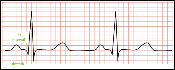
PR interval
- The AV node delays the action potential reaching the ventricles allowing the atria to contract before the ventricles
- This delay can be seen on an ECG as a brief flat section
- It forms part of the PR interval (start of P wave to start of QRS complex)
- The only way the depolarisation can spread from the atria to the ventricles is through the AV node.
- This is because there is an insulating layer at the bottom of the atria called the annulus fibrosus which prevents electrical activity from spreading

Note that even though it is called the PR interval it is actually from the start of the P wave to the start of the Q wave.
Ventricular Depolarisation
- The action potential then spreads through a network of fibres to the ventricles
- This happens very quickly
- The pattern of ventricular depolarisation is called the QRS complex
- At the same time, the atria are repolarising which can't be seen on the ECG (obscured by the QRS complex)

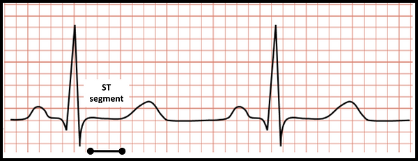
ST Segment
- There is then a brief period of net zero electrical activity
- This is a time between depolarisation and repolarisation
- This is called the ST segment

T-Wave
- Having depolarised and contracted, the ventricles move onto repolarising
- Moving ions against a concentration gradient requires energy: repolarisation takes a little bit longer than depolarisation
- This should appear as an upward deflection
- The deflection is called a T wave

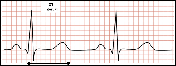
QT Interval
- So far, we have considered ventricular depolarisation and ventricular repolarisation as separate entities
- There is scope to combine the two and then consider ventricular activity as a whole
- We use the QT interval to do this
- This is often corrected for heart rate (giving us a QTc measurement) by using a mathematical formula

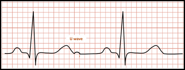
U-Wave
- Occasionally, the repolarisation process may not be completed in one smooth wave
- There may be a second phase to repolarisation (the papillary muscles and Purkinje fibres repolarise late)
- If this is the case, there may be a distinct upward deflection following the T wave (like the T wave but smaller)
- This is called the U wave

