Brady-arrhythmias and Blocks
Learn about the different types of heart block as well as how to differentiate between left and right bundle brach blocks
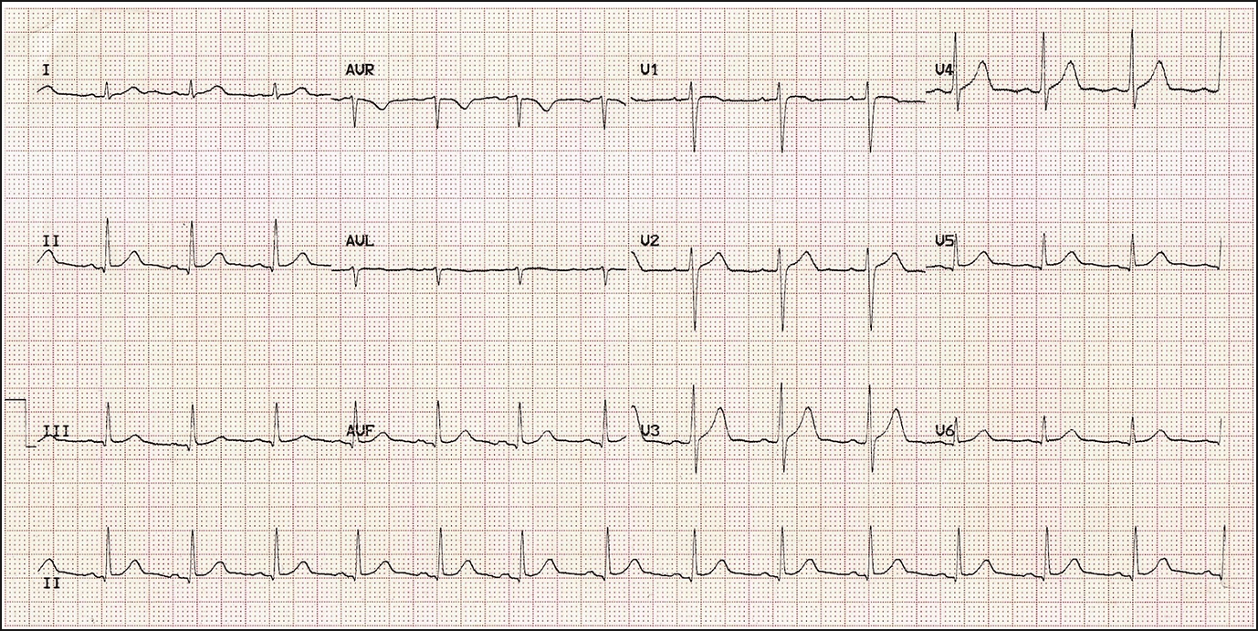
Normal Rhythm:
First Degree Heart Block:
In first degree heart block there is a delay in the depolarisation going through the AV node.
The PR interval is elongated and is longer than 200ms (5 small squares).
Sometimes if the PR elongation is longer than this the P-wave can become buried in the T-wave of the previous beat.

Second Degree AV Block Mobitz 1:
This is also known as Wenkebach phenomenon. It is caused by functional suppression of the AV node e.g. by drugs.
Here the PR interval gets progressively longer until a QRS complex is actually skipped out.
If you look at the rhythm strip after every 4th beat the next QRS is dropped.

Second Degree AV Block Mobitz 2:
Here the block occurs below the AV node, in the His Purkinje system and is due to structural damage mostly e.g. infarction
The PR interval is constant but then one of the p waves will fail to conduct (indicated by the arrows). Patients require pacemakers.
Image taken from: http://lifeinthefastlane.com/ecg-library/basics/mobitz-2/

Second Degree AV (2:1):
This type of AV block is called High grade or 2:1 AV block because there are two p waves to every QRS. Because we can only see one PR interval before there is a p wave that fails to conduct we cannot call it either Mobitz 1 or 2 and hence we don't know if the block is at the AV node or below it.

Third Degree (complete) Heart Block:
Here the impulses from the SA node are not being conducted through to the ventricles. The AV node is firing slowly by itself causing the ventricles to contract. This is called escape rhythm.
There is no correlation between the P-waves and the QRS complexes and the rate of contraction of the ventricles is slow (roughly 40 bpm) but regular.

Right Bundle Branch Block:
There is block to conduction through the right bundle branch and hence a delay to right ventricle contraction as the signal comes through the IV septum.
The QRS here is wide as conduction takes longer. As well as this if you look at V1-3 the QRS takes on an M shape due to the delay in depolarisation on the right.
In V6 we see a wide S wave which can also be slurred into the T wave
image courtesy of http://www.gnorb.net/wp-content/uploads/2011/01/rbbb.jpg

Left Bundle Branch Block:
Here the depolarisation spreads down only the right bundle branch and then across the septum to the left side. This is because there is a block in the left bundle branch.
The QRS is wide (>120ms) and the S waves in V1-3 are very long. At the bottom of these waves is a small notch giving it a W shape.
In V6 the R wave is broad and positive. It also has an M shape. All the lateral leads (I, aVF and V5-6) normally take this form but in this ECG V5 is negative.