AB1.H1.2 +D1 +D2 Accessory Glands of GI Tract, Goblet Cells, Submucosal Gland
Accessory Glands of GI Tract:
- During development of the gut tube many glands are derived from the basic gut tube which is initially formed
- In many parts of the gut tube there are mucous secreting glandular cells which form part of the lining epithelium
- These include the single celled glands known as goblet cells
- Multicellular glands develop from downgrowths of epithelial cells from the primitive gut lining into the underlying connective tissue of the lamina propria or submucosa of the gut tube
- For example, in the mouth and oesophagus there are many small glands whose secretions lubricate and add digestive enzymes to the food
- There are also some large glands which lie outside of the gut tube but retain a connection through a duct(s) which carry the secretions into the lumen of the gut tube
- These include:
- Submandibular, sublingual and parotid glands which drain into the oral cavity. (These will be studied later, during classes on the head and neck)
- Liver
- Pancreas
Questions:
Goblet Cells:
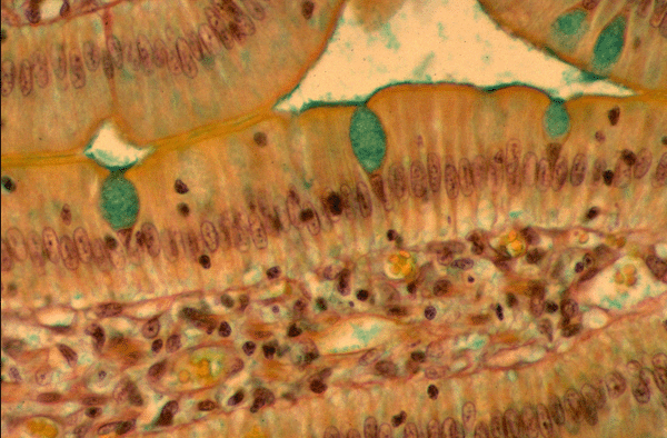
- In the micrograph shown, goblet cells are stained green
- The green staining shows the mucus secretion in storage vesicles within the single cell glands knows as goblet cells (so called because their shape is similar to a Paris goblet wine glass)
- Look closely on the apical surface of the epithelial cells to identify a thin layer of green stained mucus (derived from goblet cells)
- This mucus serves to protect the epithelium
- Goblet cells become more numerous in the large intestine, particularly at the distal end

Submucosal Gland:
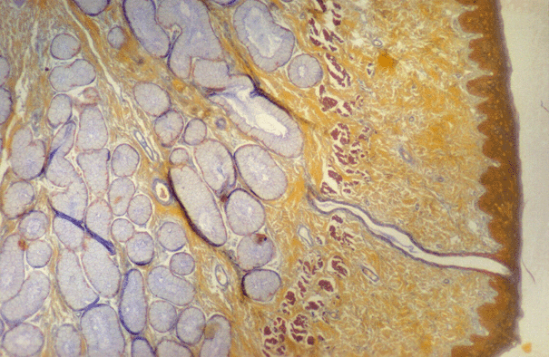
- In this micrograph identify the 3 layers of the mucous membrane of the oesophagus:- epithelium, lamina propria and muscularis mucosae
- The area of submucosa (lying deep to the mucous membrane) shown in this micrograph is occupied by a mucus secreting gland
- Notice the duct which, in life, would carry the secretions of the gland and deposit them onto the luminal surface of the epithelium

