TH1.H1.6 Veins, +D1 Peripheral Veins
Veins:
- Veins are the collecting vessels of the cardiovascular system
- They collect the blood, at virtually zero pressure, from the capillary beds and transport it back to the heart
- Veins are generally thinner walled than the arterial vessels that they are related to
- In the main, they have little or no muscle in the tunica media
- They tend to have a thick tunica adventitia which is important for supporting the walls of the veins as well as for attaching them to surrounding structures
- In arteries, the muscle of the tunica media is mainly responsible for maintaining the structural integrity of the wall
- Veins provide channels through which blood may flow
- Movement along the veins is largely a combination of external factors acting on the veins, valves in the veins controlling direction of flow, and gravity
- There is considerable variation in the structure of the walls of veins which can be related to their position in the body and/or the effects of gravity
- In general, veins superior to the heart are thinner walled than veins which lie inferior to the heart
- In the limbs, superficial veins (lying in the superficial fascia) are thicker walled than deep veins (surrounded by muscle and the deep fascia around the muscle groups)
Question:
Answer Submitted
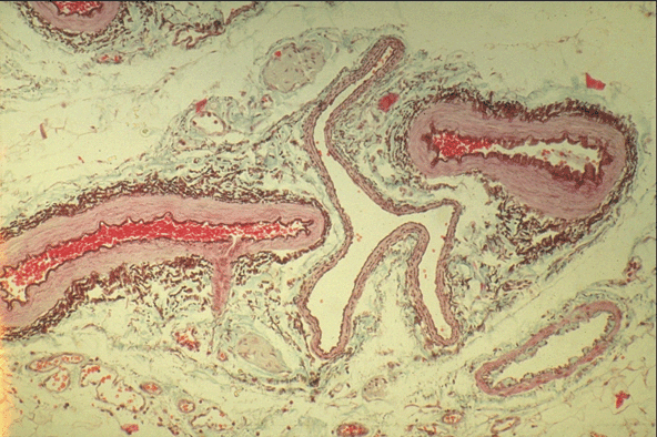
Micrograph of Peripheral Veins:
- In this micrograph there is a large vein in the centre of the picture
- To the left and right of the vein there is an artery
- Part of another vein is also shown
- Notice that the veins have a thinner wall than their corresponding arteries and that this is largely due to the thinner tunica media of the veins
- Although the wall of the vein is collapsed, you should be able to appreciate that the internal diameter of the vein is greater than that of its corresponding artery

