TH1.H1.4 +D1 Arterioles
Arterioles:
- Arterioles are small diameter vessels
- Characteristically they have only one or two layers of smooth muscle cells in the tunica media
- These vessels arise from small arteries and control the flow of blood into capillary beds
- They are richly innervated by autonomic nerves which controls the degree of contraction of the muscle in the tunica media
- Alternating contraction and relaxation of arterioles will supply small amounts of blood, in a phasic manner, into the capillary bed(s) they supply
- This phasic pattern of blood supply to capillary beds provides time for a group of red blood cells to exchange gasses across the wall of the capillaries before they are displaced by another group of red cells
- As well as this local control of blood distribution arterioles also have a collective role in the maintenance of the blood pressure
- The tone of the muscle in the tunica media of the arterioles around the body is the main factor in maintaining a stable blood pressure
- If all the muscle cells in the arterioles were to relax this would lead to an increase in the diameter, and volume, of these vessels and overall reduce the blood pressure within the arterial system
Micrograph of Arteriole:
- The arteriole is lined by an epithelium (E) forming the tunica intima of the arteriole
- The tunica media is represented by a single layer of smooth muscle cells (M)
- The tunica adventitia is indistinct in an arteriole
- In this micrograph 2 small axons (arrows) can be seen in the connective tissue adjacent to the arteriole
- It is possible that these are passing to supply the arteriolar muscle
- Arterioles have a rich innervation
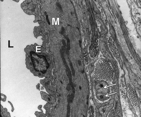
This high powered electron micrograph shows the cell types found in the wall of an arteriole, only part of the arteriole is shown (L = lumen of arteriole)
Question: