INT.H1.3 The Histological Process
The Histological Process:
-
You do not need to know the details of how tissue specimens are prepared for examination in a microscope, or the technical skills which are required for detailed histological analysis. However there are some basic points which you may find helpful.
- Histological specimens:
- Represent a thin slice, usually taken at random, through a small piece of tissue which has been chemically preserved
- Thus represents only a snapshot of what was occurring in the tissue at the time the tissue sample was taken
- Some organs have different compartments with a different structure and function
- A histological specimen may not, and usually does not, tell you everything about the tissue from which the sample was taken
- Histological sections of tissue:
- Are typically 5 microns (or less) in thickess and, usually, represent only a two dimensional view of a three dimensional structure (which has been frozen in time)
- A section in a higher or lower plane of the tissue, or from a sample taken a few seconds later, may offer different information
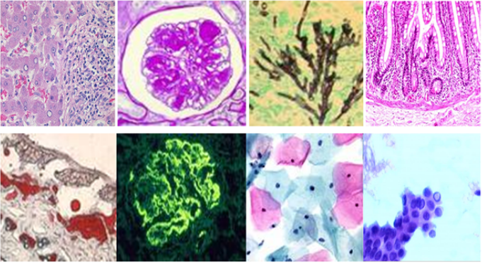
- Colour:
- With few exceptions histological sections are colourless
- Dyes are applied to section to provide some contrast to aid interpretation
- The most commonly used combination of dyes results in cell nuclei appearing blue
- That does not mean that nuclei are blue - they are not
- Study the structure not the colours