LL1.H1.7 +D1 Muscle, Tendon, Bone Connections
Muscle, Tendon, Bone connections:
- In skeletal (and smooth) muscle the contractile elements within the cytoplasm interact to cause a shortening of the fibre
- The contractile elements within the cytoplasm anchor to the inside of the cell membrane of the ends of the fibre
- The collagen fibres of the supporting connective tissue anchor into the basement membrane of the ends of the muscle fibre
- The ends of the muscle fibre are irregular to provide a greater surface area for the insertion of the collagen fibres into the basement membrane
- At the ends of each skeletal muscle, the endomysium, perimysium and epimysium converge to become continuous with the dense regular connective tissue of the tendon
- During early development, the presumptive connective tissue of the tendon is associated with the connective tissue which is destined to differentiate into cartilage/bone
- The deposition of cartilage/bone matrix around the collagen fibres of the presumptive tendon created an anchorage for the tendon
- As bones grow and increase in size, more and more of the tendon becomes incorporated into the bone matrix
- This provides an increasingly firm anchorage as the individual increase in size and the tension on the attachment site increases
- The tendinous collagen fibres that are incorporated into bone matrix are known as Sharpey's fibres
Micrograph of Dense Connective Tissue:
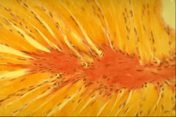
- This micrograph shows dense connective tissue (stained red) of the tendinous insertion of skeletal muscle cells of the tongue
- Note that the connective tissue (red) appears to envelope the ends of the skeletal muscle cells (orange)
- In the tongue, skeletal muscle cells are attached laterally to bone but in the midline to a band of connective tissue known as a raphe
- A similar arrangement is found in the anterior abdominal wall
- However the association between the skeletal muscle cells and the midline raphe is similar to that found where muscle cells link to a tendon

