HN3.H1.4 +D1 +D2 +D3 The Anterior Pituitary
The Anterior Pituitary:
- The anterior pituitary, or adenohypophysis, is the part of the pituitary gland that originates from the epithelium derived from the roof of the mouth
- The parenchymal cells produce 7 different hormones (growth hormone, prolactin, adrenocorticotrophic hormone, thyroid stimulating hormone, follicle stimulating hormone, luteinising hormone, melanocyte stimulating hormone)
- Cells producing each hormone are located throughout the anterior pituitary
- Mostly, each cell only produces one hormone, at least at anyone time
- The different cell populations traditionally have been distinguished by using a variety of histological staining techniques, more recently supplemented by electron microscopy examining the detailed morphology of individual cells
- However, antibodies directed against the hormones are now the preferred method for examining the frequency and location of individual hormone producing cells
- The parenchymal cells are organized into cords which are interspersed with fenestrated capillaries
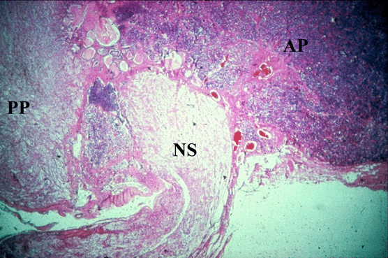
Anterior Pituitary: Low Magnification
- This low magnification mid-sagittal section of pituitary gland shows the anterior pituitary (AP) and the posterior pituitary (PP) including the neural stalk (NS)

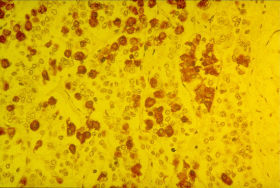
Anterior Pituitary: Growth Hormone Producing Cells
- This micrograph of anterior pituitary gland shows ‘staining’ with an antibody (labelled with brown marker) raised against growth hormone
- The distribution of growth hormone producing cells can easily be seen
- Note that they are not all clustered together in one area

Anterior Pituitary: Cords of Parenchymal Cells
- These micrographs of anterior pituitary gland show the cords of parenchymal cells and the large diameter capillaries that lie between the cords of cells
- Micrograph A is a section stained with haematoxylin and eosin which fails to male a clear distinction between the various secretory cell types found in the anterior pituitary
- Micrograph B is one of the special stains historically used to identify subpopulations of cells

