HN3.H1.1 +D1 +D2 The Thyroid Gland
The Thyroid Gland:
- The thyroid gland is located around the upper part of the trachea in the neck
- It has a very characteristic histological appearance
- The thyroid gland is composed of numerous follicles enclosed within a connective tissue capsule and attached to neighbouring structures by an additional outer fascial sheath
- Each follicle consists of a layer of simple cuboidal epithelium which encloses a mass of glycoprotein known as thyroglobulin: this is sometimes referred to as the colloid
- The thyroglobulin is produced by the cuboidal epithelial cells and deposited within the colloid where it is stored (extracellularly) and iodinated
- When the active hormones (thyroxine, triiodothyronine) are required, a small amount of thyroglobulin is endocytosed by the cuboidal epithelial cells
- Within these cells, it is modified to form the active hormones which are then released into the connective tissue that surrounds the follicle
- There, numerous fenestrated capillaries take up the hormone and transport it to its target sites
- In addition to thyroxine and tri-iodothyronine, the thyroid gland also produces the hormone calcitonin, a calcium lowering hormone
- The calcitonin producing cells are known as "C" cells (clear cells), or parafollicular cells
- These are large cells which are located in the walls of the thyroid follicles as widely spread but individual cells
- They do not abut onto the central thyroglobulin core of the follicles, or deposit any product into the core
- Calcitonin, is released directly into the connective tissue around the follicles and then passes into capillaries for distribution
Thyroid Gland: Low Magnification
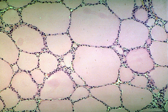
- In this low magnification micrograph of thyroid gland note the numerous thyroid follicles
- Each follicle consists of a layer of simple cuboidal epithelium surrounding a central mass of thyroglobulin (also known as colloid)

Thyroid Gland: High Magnification
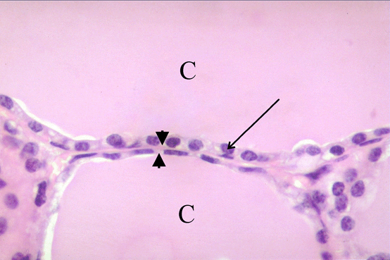
- In this high magnification micrograph identify the simple cuboidal epithelium (arrow) that surrounds a thyroid follicle and the colloid (C)
- The blood capillaries that hormone is distributed through are located in the connective tissue areas that lie between follicles (arrowheads)

Question:
Answer Submitted
