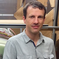
PhD, MInstP
Senior Research Fellow
- About
-
- Email Address
- l.broche@abdn.ac.uk
- Office Address
Biomedical Physics Building Room F10 Foresterhill
- School/Department
- School of Medicine, Medical Sciences and Nutrition
Latest Publications
Field cycling imaging to characterise breast cancer at low and ultra-low magnetic fields below 0.2 T
Communications Medicine, vol. 4, no. 1, pp. 221Contributions to Journals: Articles- [ONLINE] DOI: https://doi.org/10.1038/s43856-024-00644-2
Markers of low field NMR relaxation features of tissues
Scientific Reports, vol. 14, no. 1, 24901Contributions to Journals: ArticlesField-Cycling MRI for Identifying Minor Ischemic Stroke Below 0.2 T
Radiology, vol. 312, no. 2, e232972Contributions to Journals: ArticlesOpen-source magnetic resonance imaging: Improving access, science, and education through global collaboration
NMR in Biomedicine, vol. 37, no. 7, e5052Contributions to Journals: ArticlesField-Cycling Magnetic Resonance Imaging for identifying Minor Ischemic Stroke below 0.2 tesla
RadiologyContributions to Journals: Articles
- Research
-
Research Overview
I am currently leading the Fast Field-Cycling group at the University of Aberdeen, which is world-leading in the development of large-band, field-cycling imaging scanners. We are currently developing Field-Cycling Imaging (FCI), a new imaging technology derived from MRI that has the unique ability to measure the dynamics of water and lipid molecules non-invasively. This provides unique insights on the pathological remodelling of tissues during the progression of diseases, with exciting applications in medicine. FCI opens access to a new domain of medical research that remains to be explored.
I am currently conducting clinical research showing that FCI can detect stroke, breast cancer, brain glioma, liver fibrosis and osteoarthritis, amongst other pathologies. My research encompasses many disciplines such as electronic engineering, spin physics, biophysics, physiology, cell biology, system engineering or electromagnetism, and my current research direction focuses on three research topics:
- discovering the medical applications of FCI, incuding the biological mechanisms underlying the FCI image contrast
- technology developments of FCI
- dissemination of FFC imaging using open-source hardware
Research Areas

Engineering

Research Specialisms
- Biomedical Engineering
- Diagnostic Imaging
- Systems Engineering
- Medical Physics
- Electromagnetism
Our research specialisms are based on the Higher Education Classification of Subjects (HECoS) which is HESA open data, published under the Creative Commons Attribution 4.0 International licence.
Current Research
Clinical applications of Field-Cycling Imaging
Field-Cycling Imaging (FCI) is a unique imaging technique that has the ability to quantify the motion of water and lipids in vivo, non invasively and using low and safe magnetic fields. Water naturally diffuses through the body and interacts with all its components, hence water motion is sensitive to the pathological tissue remodelling that occur as diseases progress. Using FCI, it is possible to detect and measure these changes, and therefore to detect and follow the progression of certain diseases.
Our pilot studies show excellent results in several applications:
Characterisation of brain stroke in vivo
The management of stroke has increasingly focused on early identification, early scanning and thrombolysis and/or clot retrieval. These techniques rely heavily on imaging of the stroke but there are practical and interpretational limitations to current imaging modalities for early diagnosis of ischaemic stroke.
Field-Cycling Imaging (FCI) uses low magnetic fields to extract non-invasively information on the molecular dynamics of tissues that is not accessible by any other imaging modalities. Our PUFFINS pilot study shows great potential to identify ‘what is going on’ at the molecular level after a brain infarct and to give information on the composition of intra-arterial thrombus, which might aid choice of treatments for acute strokes. Other potential benefits of FCI are foreseen for patients with small vessel disease and amyloid deposition, not to mention other brain diseases such as vasculitis, tumour and multiple sclerosis.
Detection of osteoarthritis using the quadrupolar signal
Osteoarthritis (OA) is the most prevalent joint disorder and cause of disability in the United Kingdom with an estimated 8.5 million persons suffering from joint pain due to OA, and it is a major cause of disability in the world. Multiple risk factors appear to be involved in the onset and progression of OA, including age, genetics, gender, overuse, trauma and obesity, even though none of these have been identified as a clear cause of the disease. There are currently no pharmacological interventions available to patients for modifying the underlying disease but early patient management can help to slow down its progression. it is therefore crutial to detect OA before irreversible damages develop.
The underlying pathophysiology of OA has been extensively studied and recent research identifying the importance of matrix-degrading enzymes, chondrocyte hypertrophy and apoptosis, subchondral bone metabolism, cytokines and inflammation has identified a number of potential targets for disease modifying agents. Interest in developing potential therapeutic agents has highlighted the need for biomarkers of disease progression with imaging biomarkers currently appearing to offer the best prospect.
A previous study of FCI on excised samples of OA cartilage from hip replacement showed promising results: it appeared that this technique can offer degradation-sensitive contrast using a particular signal, the quadrupolar signal. A more extensive study is being undertaken at the moment in collaboration with Mr G.P. Ashcroft and Prof R. Aspden in order to assess this technique.
Characterisation of fibrin clots and applications to thrombosis
Fibrin is one of the main constituents of blood clots. It is derived from fibrinogen, a long, narrow and heavy protein (340 kDa), under the action of thrombin.
Fibrin protein are insoluble and aggregate into long filaments that can cross-link to form a rigid gel structure. The size and diameter of these filaments, together with the amount of cross-linking, can be controled more or less independantly during the clotting process by several paremeters such as the concentration of calcium ions or the content of factor XIII and RS283 proteins.
The rigidity of the clot is an important parameter for the treatment of deep vein thrombosis (DVT), a pathology that may develop in several diseases or conditions such as cast ankle, high saturated fat diet or drug abuse. DVT is treated by thrombolysis when the conditions allow, but response to treatment varies greatly due to the nature of the clot.
FFC NMR studies of fibrin clots have showed that field-cycling can detect fibrin quantitatively, with possible applications to DVT characterisation. A study has been initiated on DVT patients in parternship with the NHS at the Aberdeen Royal Infirmary to assess whether FCI can predict the response to thrombolitic treatment.
An in-vitro study of fibrin systems is also being developed in partnership with Dr N. Mutch and Dr C. Whyte that aims to detect differences in clot structure due to the effect of polyphosphate chains during the clot.
Detection of tumours at low magnetic fields
one of the parameters of interest that is measured by an MRI scan is the transverse relaxation time of spins, also called T1. This parameteris known to vary greatly between tissues at low magnetic field (typically below 0.05T) but much less at high fields. This is part of the reason why contrast agents are needed for tumour detection on conventional MRI clinical scanners.
Unfortunately, low magnetic fields also come with low image resolution and long scan time. This is the reason why MRI scanners are operating at ever increasing fields.
FCI offers a compromise between high contrast and high resolution that cannot be reached by conventional fixed-field MRI scanners. This opens new avenues for contrast-based studies with potential applications in many fields of medicine.
One particular area of interest is breast cancer: breast tumours are commonly detected by analysis of contrast in MRI scans but may be difficult to assess if their extent is restricted to a few millimeters. A study has been initiated by Dr Lionel Broche in collaboration with Prof S. Heys, Dr I. Miller, Dr T. Gagliardi, Mr G.P. Ashcroft, Dr D. Boddie and Dr S. Dundas to assess FFC MRI in the context of breast cancer.
Funding and Grants
See my ORCID profile for up-to-date details.
- Teaching
-
Teaching Responsibilities
Dr Lionel Broche teaches the physics of Fast Field-Cycling Magnetic Resonance Imaging on the MSc programmes in Medical Physics, Medical Imaging, and Medical Physics Computing.
- Publications
-
Page 9 of 11 Results 81 to 90 of 110
Fast field-cycling magnetic resonance imaging: a new imaging modality
European Congress of Radiology (2015), B-0315Contributions to Journals: Abstracts- [ONLINE] DOI: https://doi.org/10.1007/s13244-015-0387-z
Rapid field-cycling MRI using fast spin-echo
Magnetic Resonance in Medicine, vol. 73, no. 3, pp. 1120-1124Contributions to Journals: Articles- [ONLINE] DOI: https://doi.org/10.1002/mrm.25233
- [OPEN ACCESS] http://aura.abdn.ac.uk/bitstream/2164/5580/1/RapidFFCMRIusingFSE.pdf
Calibration of a Fast Field Cycling Relaxometer for Ultra‐Low Field Measurements of T1‐Dispersion
Annual Scientific Meeting of ISMRM British Chapter (2014), pp. P1BContributions to Conferences: AbstractsClinical applications of Fast Field‐Cycling techniques
Annual Scientific Meeting of ISMRM British Chapter (2014), pp. PP1AContributions to Conferences: AbstractsDevelopment of a voltage‐tuneable RF coil to enable double resonance experiments with Fast Field‐Cycling MRI
Annual Scientific Meeting of ISMRM British Chapter (2014), pp. P1CContributions to Conferences: AbstractsField-Cycling MRI: a New Imaging Modality?
8th European Conference on Medical Physics (2014), pp. e8-e9Contributions to Conferences: Abstracts- [ONLINE] DOI: https://doi.org/10.1016/j.ejmp.2014.07.037
A varactor-tuned RF coil for nuclear quadrupole double resonance with Fast Field-Cycling MRI
AMPERE NMR School (2014), pp. P74Contributions to Conferences: AbstractsCalibration of a Fast Field Cycling Relaxometer for Ultra-Low Field Measurements of T1-Dispersion
AMPERE NMR School (2014), pp. P92Contributions to Conferences: AbstractsTechniques and Bio-Medical Applications of Field-Cycling Magnetic Resonance
AMPERE NMR School (2014), pp. 21Contributions to Conferences: AbstractsFast field-cycling NMR of cartilage: a way toward molecular imaging
World Congress of the Osteoarthritis-Research-Society-International (OARSI) (2014), pp. S66-S67Contributions to Journals: Abstracts
